Time-lapse Shadow Image Analysis
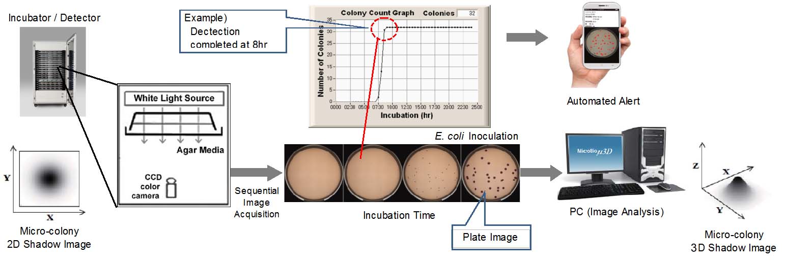
This system monitors spread or pour plate in microscopic level, detects and counts micro-colonies, drawing colony count graph automatically, while incubating the plate. The system automates the traditional agar media culture method as it is.
Advantage
- Just automating the traditional culture method
- Easy to use (Place agar plates and start a test.)
- Microbial detection is rapid and real time. (It detects micro-colonies.)
- Colony count is accurate.
- Spread or pour plate can be tested.
- Membrane filter method can be automated for rapid microbial detection as it is.
- Agar media performance can be evaluated by using a graph.
- Plate images sampled in every 30 minutes are saved in digital storage.
- The colony developing process can be verified by reviewing stored image data, going back in time, even in the middle of testing or after the test is completed.
- Agar media performance can be evaluated precisely by using a graph.
Not only by a visual inspection, but also by a colony count graph, the microbial growth will be verified so that an agar media performance can be evaluated easier and more precisely.
Time-lapse Shadow Image Analysis was invented by the company’s founder, Dr. Hiroyuki Ogawa, Ph.D. The MicroBio μ3D Test System is the first and only fully automated microbiological test system that addresses all microbial test applications and fits with current regulatory practices, accelerating to obtain the precise test results since the system is just automating the traditional culture method in every aspect.
Hardware
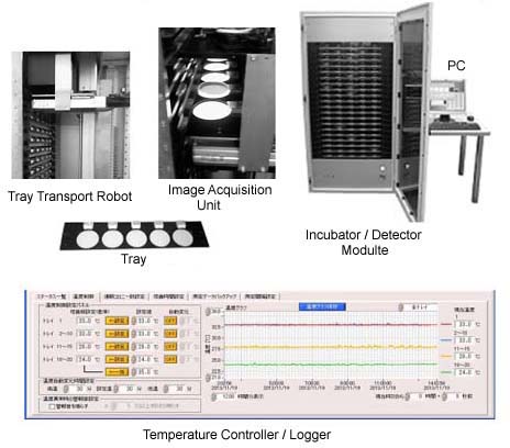
MicroBio µ3D AutoScanner system based on Time-Lapse Shadow Image Analysis (TISA) is consisted of incubator module, tray handling robot, image acquisiton unit and a PC for test control and image analysis.
The incubator module accomodates 20 trays on the shelves that are made out of heater plates.
Four different temperature can be assigned to four groups of selves, if necessary. Typical application is to set 20℃ to lower 20 trays and 35℃ to upper 20trays, for mold detection and for bacteria detection, respectively. The temperature cycle function is also available.
Basic Performance
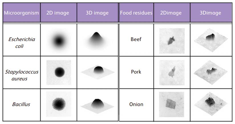
Microbial Colony and Food Fragment
Microbial colony exhibits round shape in 2D shadow and conical shape in 3D shadow image.
In case of food fragment, the shpe is irregular.
Time-lapse Shadow Image Analysis (TSIA) versus Visual Inspection
The detection and count result of both method were the same.
The following was how the verification was conducted.
- The pour Desoxycholate agar plate was prepared to produce about 250 colonies of E. coli.
- At every one hour, count result of the same plate was obtained by TSIA system and visual inspection.
- For visual inspection, 5 times magnifier lens was used.
- Incubation was continued until 16hr.
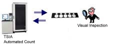
The count result was 254 for both methods at 16hr.
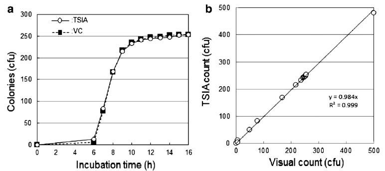
(a) TSIA and Visual colony count in chronological order (b) Correlation between both methods, n=15
Thesis
There are the following papers on this method.
- Journal of Microbiological Methods. 2012 Dec;91(3):420-8. doi: 10.1016/j.mimet.2012.09.028. Epub 2012 Oct 22. Noise-free accurate count of microbial colonies by time-lapse shadow image analysis
- Scientific Reports. 2015 May,Rapid and retrievable recording of big data of time-lapse 3D shadow images of microbial colonies
- Biocontrol Science. 2015 Dec, Accurate Enumeration of Aspergillus brasiliensis in Hair Color and Mascara by Time-Lapse Shadow Image Analysis
Related Page
- Agar media Performance:
Evaluation method of agar culture media - Precise evaluation of agar culture media is achieved by MicroBio µ3D AutoScanner with colony enumeration graph and histogram